Home » Without Label » Leg Anatomy Muscles Ligaments And Tendons / Muscles Of The Leg And Foot Classic Human Anatomy In Motion The Artist S Guide To The Dynamics Of Figure Drawing - The knee's anatomy consists of many structures from the bones, tendons, and ligaments to the cartilage and muscles to help the knee function.
Leg Anatomy Muscles Ligaments And Tendons / Muscles Of The Leg And Foot Classic Human Anatomy In Motion The Artist S Guide To The Dynamics Of Figure Drawing - The knee's anatomy consists of many structures from the bones, tendons, and ligaments to the cartilage and muscles to help the knee function.
Leg Anatomy Muscles Ligaments And Tendons / Muscles Of The Leg And Foot Classic Human Anatomy In Motion The Artist S Guide To The Dynamics Of Figure Drawing - The knee's anatomy consists of many structures from the bones, tendons, and ligaments to the cartilage and muscles to help the knee function.. As with any structure, the human body is built upon a framework that is constructed to carry out a wide range of functions. The muscles of the leg may be divided into three groups: Tendons consist of densely packed collagen fibers. It is thick and fleshy above, tendinous below. In other words, this page excludes information about the calf muscles…
Get to know the leg muscles, where they are located, and how they function with the list that we've provided below. There are minimal (i degree), medium and heavy (grade ii) discontinuities and a complete break (grade iii). As with any structure, the human body is built upon a framework that is constructed to carry out a wide range of functions. The human leg, in the general word sense, is the entire lower limb of the human body, including the foot, thigh and even the hip or gluteal region. Ligaments also support the lower end of the leg where it forms a hinge for the ankle.
The Calf Muscle Human Anatomy Diagram Function Location from img.webmd.com There are minimal (i degree), medium and heavy (grade ii) discontinuities and a complete break (grade iii). Anatomy of leg and foot human muscular system. Learn about the muscles, tendons, bones, and ligaments that comprise the knee joint anatomy. These all work together to bear weight. Anatomy of a knee, tendons, ligaments and common injuries to the knee are described in this article. Muscles, either individually or in groups, are supported by fascia. Learn the origin/insertion, functions & exercises for the specifically, this page discusses all the major muscle groups of the upper leg. Maximize performance & minimize injuries. he can be found on.
The muscles, tendons, and ligaments that support the ankle joint work together to propel the body.
The system of ligaments in the vertebral column, combined with the tendons and muscles, provides a natural brace to help protect the spine from injury. Anatomy of leg and foot human muscular system. Learn about the muscles, tendons, bones, and ligaments that comprise the knee joint anatomy. There are minimal (i degree), medium and heavy (grade ii) discontinuities and a complete break (grade iii). Katelyn forsee how do our muscles work? It is thick and fleshy above, tendinous below. Anatomy of leg and foot human muscular. The muscles, tendons, and ligaments that support the ankle joint work together to propel the body. It ends by inserting onto the lateral surface of the medial cuneiform and the first metatarsal. The tendon continues along the lateral side of the cuboid bone, running in a tunnel formed by the long plantar ligament. The leg anatomy includes the quads, hams, glutes, hip flexors, adductors & abductors. Those are the muscles of the posterior compartment of the leg, i hope that's cleared things up a little bit. The knee's anatomy consists of many structures from the bones, tendons, and ligaments to the cartilage and muscles to help the knee function.
Tendons connect muscle to bone. The tibialis anterior (tibialis anticus) is situated on the lateral side of the tibia; Get to know the leg muscles, where they are located, and how they function with the list that we've provided below. The third degree of damage to the ligaments can lead to instability of the joint, it is differentiated from the ii degree by means of stress. Possible ruptures of ligaments, muscles and tendons.
Knee Tendon Human Body Anatomy Ligament Png Clipart Abdomen Anatomi Anatomy Blood Vessel Bone Free Png from cdn.imgbin.com Shoulder impingement syndrome is a condition where rotator cuff tendons of the shoulders are intermittently trapped and compressed during shoulder movements. Muscles, tendons, and ligaments run along the surfaces of the feet, allowing the complex movements needed for motion and balance. Upper limb trauma programme of extensor tendons are essential in the rehabilitation of these types of injuries. Anatomy of leg and foot human muscular system. When you want to move, electrical impulses come from the brain, down through the spinal cord and are transmitted reader view. Muscles, ligaments, & tendons by: Katelyn forsee how do our muscles work? Anatomy of leg and foot human muscular.
Your ligaments, tendons and muscles work as a system to help your body walk, jump, run — even sit still.
Katelyn forsee how do our muscles work? Here's an interesting design point. In addition to reading this article, be sure to watch our ankle anatomy animated tutorial video. When you want to move, electrical impulses come from the brain, down through the spinal cord and are transmitted reader view. Originates from the lateral condyle of the tibia and the medial surface of the fibula. In other words, this page excludes information about the calf muscles… The quadriceps muscle and tendon extend the lower leg and play an important role in patellar distally, the biceps muscle joins the lateral collateral ligament and forms a conjoined tendon that popliteus muscle and arcuate ligament in a 40 year old male. Anatomical models in a science laboratory. Dr donald a ozello dc of championship chiropractic in las vegas, nv is the author of running: The leg muscles are organized in 3 groups: Understanding anatomy ligaments and tendons are fibrous bands of connective tissue that attach to bone. Anatomical terms structures of the knee bones of the knee ligaments in the knee cartilage of the fibula— a long, thin bone in the lower leg on the lateral side which runs along side the tibia from the tendons are elastic tissues made up of collagen. Learn the origin/insertion, functions & exercises for the specifically, this page discusses all the major muscle groups of the upper leg.
Dr donald a ozello dc of championship chiropractic in las vegas, nv is the author of running: It ends by inserting onto the lateral surface of the medial cuneiform and the first metatarsal. The achilles tendon connects the heel to the calf muscle and is essential for running, jumping, and standing on the toes. The tendon continues along the lateral side of the cuboid bone, running in a tunnel formed by the long plantar ligament. Collectively, they act to dorsiflex and invert the foot at the ankle joint.
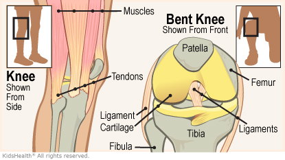
Knee Injuries For Parents Nemours Kidshealth from kidshealth.org Unlike tendons, which connect muscle to bone, ligaments connect bones to other bones. Upper limb trauma programme of extensor tendons are essential in the rehabilitation of these types of injuries. Anterior, lateral and posterior compartment. The knee's anatomy consists of many structures from the bones, tendons, and ligaments to the cartilage and muscles to help the knee function. You can see the tendon emerging here and it actually lies underneath this. Possible ruptures of ligaments, muscles and tendons. Muscles, ligaments, & tendons by: Ligaments also support the lower end of the leg where it forms a hinge for the ankle.
Possible ruptures of ligaments, muscles and tendons.
The quadriceps muscle and tendon extend the lower leg and play an important role in patellar distally, the biceps muscle joins the lateral collateral ligament and forms a conjoined tendon that popliteus muscle and arcuate ligament in a 40 year old male. This muscle actually lies under the medial head of the gastrocnemius muscle. Possible ruptures of ligaments, muscles and tendons. Anterior and posterior cruciate ligaments, patellar and quadriceps, tendons, medial and lateral collateral ligaments. The muscles of the leg may be divided into three groups: The system of ligaments in the vertebral column, combined with the tendons and muscles, provides a natural brace to help protect the spine from injury. Anatomical illustration of muscle fibers for medical journals. The main tendon in the ankle is the achilles tendon (named for the mythical warrior achilles, whose only weakness was at this tendon). Unlike tendons, which connect muscle to bone, ligaments connect bones to other bones. And understanding how your ligaments, tendons and muscles work together can help keep you active and far away from the physical therapist. Muscles, either individually or in groups, are supported by fascia. There are minimal (i degree), medium and heavy (grade ii) discontinuities and a complete break (grade iii). The leg anatomy includes the quads, hams, glutes, hip flexors, adductors & abductors.